Science Pool

Cyprotex

Recent Posts

ICH M12 2024 vs FDA 2020 DDI Guidance - Exploring the Differences

Posted by Cyprotex on Jun 19, 2024 6:18:50 PM

ICH M12 2024 vs FDA 2020 DDI Guidance. Exploring the Differences.

Download our handy comparison summary

The new ICH M12 guideline on drug interaction studies is welcomed by industry as it provides a harmonized approach which is expected to be implemented by the major regulatory authorities such as the US Food and Drug Administration (FDA), European Medicines Agency (EMA) and the Japanese Pharmaceuticals and Medical Devices Agency (PMDA) and presumably replace their respective existing guidance. In this blog, we explore the differences between the now finalized ICH M12 guideline adopted in May 2024 (draft for comment released 2022) and the previous US FDA 2020 guidance released in January 2020 with a focus on the in vitro assays.

Before we launch into the individual assays, one general observation in the new ICH M12 guideline is the replacement of the term ‘victim’ to ‘object’ to denote an investigational drug (usually a substrate) which is affected by a concomitant drug, and the replacement of the term ‘perpetrator’ to ‘precipitant’ to denote an investigational drug (typically an inhibitor or inducer) which affects a concomitant drug. This amendment only appeared in the final version of the ICH M12 guideline as it was one of the comments from review of the draft version.

Reaction Phenotyping

If we consider reaction phenotyping initially, the differences between the new ICH M12 and the FDA 2020 guidance are relatively minimal. Although the ICH M12 refers to all the enzymes mentioned in the FDA guidance, it also refers to some additional Phase II enzymes which may need to be evaluated including glutathione S-transferases (GSTs) and N-acetyl transferases. Both the ICH M12 and the FDA guidance have the same cut-off of ≥25% of total elimination identified using in vitro studies (reaction phenotyping and metabolite identification) and human mass balance data for determining if the enzyme needs further investigation in a clinical study. One key difference is that the FDA guidance recommends two methods should be used for reaction phenotyping – the first method using selective inhibitors in the presence of human liver microsome or hepatocytes and the second method using human recombinant enzymes. In the ICH M12 guidelines, only one of these methods is required.

Both the ICH M12 guideline and the FDA guidance have recommendations for evaluating metabolites in reaction phenotyping studies if the exposure levels of the metabolite results in clinically relevant changes in efficacy or safety.

Enzyme Inhibition

Moving onto enzyme inhibition, both the ICH M12 and FDA guidance recommend evaluating the main seven CYP isoforms for reversible and time dependent inhibition. For reversible inhibition, the cut-off for determining if a clinical study is required is the same for both the ICH M12 and FDA. However, for time dependent inhibition, 5 x Cmax,u is used in the calculation in the ICH M12 whereas a higher safety factor of 50 x Cmax,u was used in the 2020 FDA guidance, suggesting a less conservative approach is used in the new ICH M12 guideline.

One addition to the ICH M12 guideline, which is not specifically included in the previous FDA 2020 guidance, is the inclusion of reversible UGT inhibition. The ICH M12 recommends that UGT1A1, UGT1A4, UGT1A9, UGT2B7 and UGT2B15 inhibition should be evaluated if direct glucuronidation is one of the major elimination pathways of the investigational drug. Although it is considered an area of ongoing research, it is currently recommended in the ICH M12 that the same cut-off values should be used for UGT inhibition that is applied to CYP enzymes (i.e., Cmax,u/Ki,u<0.02).

Enzyme Induction

For enzyme induction, the FDA guidance suggested either catalytic activity or mRNA could be used to assess induction. It also suggested that immortalized hepatic cell lines may be used to determine CYP induction potential. However, the new ICH M12 primarily recommends the analysis of mRNA (with the exception of CYP2C19 where catalytic activity should be measured) and that the CYP induction assessment should be performed in human hepatocytes. The ICH M12 guideline also addresses the issue of toxicity and recommends that cell viability assessment is performed before and at the end of the incubation.

For data analysis, both the ICH M12 and FDA cover the three basic methods (fold-change, relative induction score correlation method and basic kinetic model) and align on the cut-offs for indicating if an investigational drug has the potential to induce in vivo. However, for the basic mRNA fold method in the ICH M12 guideline describes assessing test drug concentration of 50 x Cmax,u whereas the FDA only suggests testing up to 30 x Cmax,u suggesting a more conservative approach now by the ICH M12. Furthermore, in the calculations for the correlation methods and the basic kinetic model, unbound EC50 (EC50,u) is specified in the ICH M12 whereas only EC50 is referred to in the FDA guidance. Finally, the ICH M12 describes in more detail an indicative positive control response for CYP1A2, CYP2B6 and CYP3A4 of typically at least 6-fold to ensure sufficient sensitivity of system, whereas this level of detail is absent from FDA.

Transporter Substrate Identification

Both the ICH M12 guideline and the FDA DDI guidance (2020) recommend the same transporters are assessed. The method for testing and thresholds for clinical assessment are very similar between the FDA and ICH M12.

Transporter Inhibition

For transporter inhibition, the same transporters are recommended in the ICH M12 as the previous FDA guidance, however, the cut-off value for determining if a clinical study should be performed are different in a couple of instances. Firstly, for P-gp and BCRP inhibition where the investigative drug is administered by the parenteral route or if a metabolite is formed post absorption, the cut-off value in ICH M12 is an IC50 of 50 x Cmax,u, whereas in the FDA guidance it is 10x Cmax total; important to note the additional difference for what is defined as [I]. This indicates a more conservative approach by the FDA when plasma protein binding is > 80%, or by ICH M12 when plasma protein binding is < 79%. The second instance applies to the cut-off value for MATE1 and MATE2-K, which has increased to an IC50 of 50 x Cmax,u rather than 10x Cmax,u, indicating a more conservative approach by the ICH M12 for these renal transporters. The ICH M12 also specifically refers to unbound IC50 (IC50,u) in the calculations whereas the FDA guidance only references IC50. In this regard, it is important to recognize that any correction of IC50 for potential non-specific binding that might occur within a transporter inhibition assay would only be required if the assay did not incorporate a pre‑incubation step with investigational drug as standard methodology for all transporters prior to the co‑incubation with fresh investigational drug solution and probe substrate. With such standard methodologies, the inclusion of the pre‑incubation step would be anticipated to mask any non-specific binding sites therefore the co‑incubated concentrations of investigational drug would be nominal for IC50 fitting purposes, i.e. IC50 = IC50,u.

DOWNLOAD OUR COMPARISON SUMMARY

Get in touch

Tags: Blog, ADME/DMPK, IND Enabling Studies/Preclinical Development

Improved Predictive Power in Cardiac Risk Assessment

Posted by Cyprotex on Dec 21, 2023 3:09:38 PM

Cardiotoxicity is one of the most reported adverse effects that leads to pre-clinical and clinical drug failure. To tackle this, the International Conference of Harmonization (ICH) S7B guideline in 2005, proposed a non-clinical assessment of new drug entities using in vitro electrophysiology studies (typically hERG ion channel) and in vivo telemetry in animal models. Although these are very sensitive approaches, they may have also led to unwarranted drug attrition of many potentially valuable therapeutics due to the low specificity nature of the assays.

More recently, the Comprehensive in vitro Proarrhythmia Assay (CiPA) initiative was proposed by experts in the field and was established to move safety pharmacology towards in silico and in vitro approaches utilising new and emerging technologies such as stem-cell-derived cardiomyocytes. Building on this new safety paradigm, colleagues from Evotec and Cyprotex have worked collaboratively to develop a non-clinical model using cutting-edge techniques with improved predictive power to de-risk cardiotoxicity in early drug discovery. We have presented the output of this work in a research article titled In-depth mechanistic analysis including high-throughput RNA sequencing in the prediction of functional and structural cardiotoxicants using hiPSC cardiomyocytes” published in a recent edition of Expert Opinion on Drug Metabolism and Toxicology.

In this article, we describe the use of human-induced pluripotent stem cell-derived cardiomyocytes (hiPSC-CMs) as an in vitro model system together with three high-throughput technologies incorporating structural assays (high-content imaging, HCI), functional assays (Ca2+ transience, CaT) and high-throughput RNA-sequencing (ScreenSeq) for the pre-clinical risk assessment of novel compounds. The transcriptional responses of hiPSC-CMs to 24 h treatment with 33 cardiotoxicants (12 structural cardiotoxicants, 14 functional cardiotoxicants, 7 structural/functional cardiotoxicants) and 9 non-cardiotoxicants of mixed therapeutic indications were investigated and compound-induced differential gene expression (DEG) was calculated in comparison with vehicle treated controls. Likewise, the hiPSC-CMs responses to six structural readouts (cell count, cellular ATP, mitochondrial mass, mitochondrial membrane potential, calcium content, DNA structure and nuclear size) and four functional readouts (amplitude, frequency, peak width and decay time) were analysed. In summary, hiPSC-CMs recapitulated expected structural and functional toxicity mechanisms, validating their use as in vitro model system to detect and characterize modes of toxicity. ScreenSeq identified several molecular mechanisms of toxicity such as alterations in cardiac pathways, genotoxicity, ER stress and mitochondrial toxicity. Together, HCI, CaT and ScreenSeq provided the best cardiotoxicity prediction metrics (10x Cmax: 100% specificity, 82% sensitivity, 86% accuracy; 25x Cmax: 89% specificity, 91% sensi-tivity, 90% accuracy).

This study not only provides invaluable cardiotoxic mechanistic information of the drugs tested, but it also demonstrates the potential of this mechanism-driven risk assessment approach in predicting drug-induced cardiotoxicity in hiPSC-CMs.

Read the paper.

Read more about our Cardiotox Screen assay consisting of both a functional assay (examining the mechanical function of the cardiomyocytes) and a structural assay (assessing morphological changes and loss of viability).

In addition, discover more about our transcriptomics offerings here.

Find out more:

Contact Us

Tags: Toxicology, Articles & Whitepapers, Blog, Toxicology & Safety

The New Cyprotex e-Store - ADME-Tox Services Now Available Online!

Posted by Cyprotex on Oct 10, 2023 1:38:12 PM

Rapid turnaround is essential for ADME-Tox services, especially during drug discovery. However, turnaround time for the assay isn’t the only consideration. The time required to prepare the quotation may delay the scheduling of the assay and, in turn, the study start date.

The new Cyprotex e-Store provides a solution to this. Registered users of the e-Store are able to view instant pricing for a comprehensive range of ADME-Tox services. As well as having access to detailed protocols and the latest editions of our educational guides, users also benefit from special offers only available through the e-Store.

Online ordering is simple. Previous orders can be tracked through the e-Store and it is easy to re-order your favourite assays. All of this information can be accessed outside business hours. A range of payment options are available on the e-Store including credit card, existing purchase order or invoice. These features allow services to be ordered and scheduled the same day so saving you time.

Despite this automated approach, you are still supported by a dedicated scientific study manager and account manager who are on-hand to guide you at all stages of your study.

Want to learn more?

Explore the Cyprotex e-Store

Tags: Blog, ADME/DMPK

Mitigating False Negatives in P-gp & BCRP Substrate Identification

Posted by Cyprotex on Sep 27, 2023 11:00:00 AM

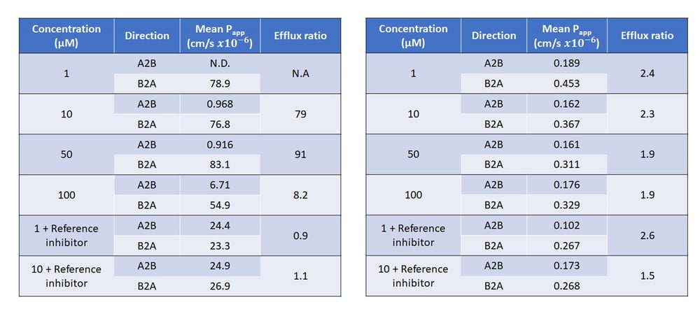
The industry gold-standard in vitro cell based test system for studying Breast Cancer Resistance Protein (BCRP) or P-glycoprotein (P-gp) transporters is the polarized cell monolayer system using either Caco-2 cells or MDCK-MDR1 cells (Elsby et al., 2022), grown on semi permeable membrane inserts to form a brush border membrane barrier separating two experimental compartments (apical and basolateral) of equivalent pH (7.4). Apparent permeability (Papp, cm/s x10-6) of the test article is determined in the apical to basolateral (A-B) and the basolateral to apical (B-A) direction and from this an efflux ratio is calculated (B-A Papp/A-B Papp). An efflux ratio greater than 2 which is reduced by greater than 50% with a corresponding reduction in B-A Papp in the presence of a reference inhibitor (Atkinson et al., 2022) indicates the test article is a substrate of the efflux transporter being investigated. The bidirectional Papp values and subsequent efflux ratios of the known P-gp substrate quinidine, determined in polarized MDCK-MDR1 cell monolayers across a concentration range is summarized in Table 1 (left) and demonstrates that in the MDCK-MDR1 polarized monolayer test system quinidine has been determined to be a substrate of the P-gp transporter.

False Negatives in PgP BSEP Fig 1

Table 1: Mean Papp values and corresponding efflux ratios of quinidine (left) and N-methyl quinidine (right) across MDCK-MDR1 monolayer cells in the presence and absence of reference inhibitor cyclosporin A

Despite polarized monolayers being the industry gold standard test system for studying P-gp and BCRP interactions, there are limitations and risk of inconclusive results and false negatives if the test article being studied exhibits low Papp indicating inherently low passive membrane permeability. This is due to the physico-chemical characteristics of the test article limiting cellular entry required to access the efflux transporter on the apical membrane. This is true for the quinidine oxidative metabolite, N-methyl quinidine (NMQ). The presence of the methyl group results in lower lipophilicity and therefore passive permeability without diminishing its ability to bind P-gp. As summarized in Table 1 (right) bidirectional Papp values for the more polar NMQ are very low and although efflux ratios determined are around two, a 50% reduction in efflux ratio with corresponding decrease in B-A Papp is not observed in the presence of inhibitor therefore the result is inconclusive at best, or at worst could be interpreted as not a substrate of the P-gp transporter.

Where assessment of a test article in polarized monolayer cells indicates a compound has inherently low passive permeability, membrane vesicles have the membrane orientated “inside out”, this results in the intracellular binding site of the transporter being positioned on the outside of the vesicle (outwardly facing), therefore compounds do not need to cross a membrane in order to interact with the transporter. As such for efflux transporters such as P-gp or BCRP, substrates will be actively taken up into the vesicle and, due to poor permeability, will remain inside the vesicle for quantification. As demonstrated in Table 2, using P-gp membrane vesicles as an in vitro test system for P-gp substrate identification, NMQ is clearly demonstrated to be, and subsequently correctly identified as, a substrate of the P-gp transporter.

False Negatives in PgP BSEP Fig 2

Table 2: Mean uptake rate and corresponding uptake ratios of N-methyl quinidine using P-gp expressing membrane vesicles in the presence of ATP or AMP in the absence and presence of the reference inhibitor cyclosporin A. Figure: Mean uptake rate of N-methyl quinidine into P-gp expressing membrane vesicles in the presence of ATP or AMP in the absence and presence of the reference inhibitor (I) cyclosporin A

Furthermore, if data from P-gp or BCRP substrate identification studies in polarized cell monolayers indicate a compound has inherently low passive permeability then any P-gp or BCRP inhibition data generated in the same in vitro test system should also be scrutinized and a follow up inhibition study in membrane vesicles considered, particularly if no inhibition was observed in the cells.

Whilst membrane vesicles have their advantages over polarized cell monolayers as a test system to identify P-gp or BCRP substrates that are poorly permeable, it is not advisable to utilize membrane vesicles as a first-choice test system. This is because for lipophilic test articles such as quinidine, sufficient passive permeability allows the substrate to move freely across the membrane and would not be trapped within the vesicle lumen, therefore there is a risk of inconclusive results and false negatives.

In summary, polarized cell monolayers are the gold standard for studying P-gp and BCRP interactions as indicated in regulatory guidances and as such should be the first choice of test system for identifying P-gp or BCRP substrates and inhibitors. However, if results indicate the compound has inherently low passive permeability then P-gp or BCRP expressing membrane vesicles should be considered as an alternative follow up in vitro test system to mitigate against the clinical implications of false negatives.


References:

Atkinson, H., Mahon-Smith, K. and Elsby, R. (2022) ‘Drug permeability and transporter assessment: Polarized Cell Lines’, The ADME Encyclopedia, pp. 401–412. doi:10.1007/978-3-030-84860-6_142.

Elsby, R. et al. (2022) Studying the right transporter at the right time: An in vitro strategy for assessing drug-drug interaction risk during drug discovery and development. Expert Opin Drug Metab Toxicol 18(10):. 619–655. doi:10.1080/17425255.2022.2132932.


Tags: Blog, ADME/DMPK